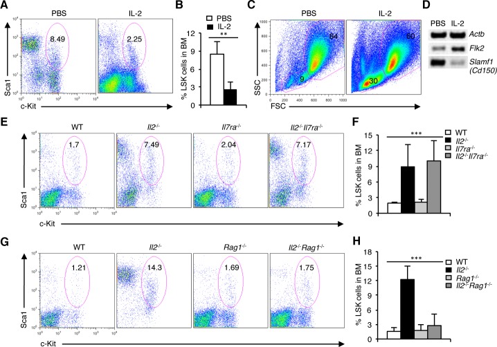
Figure 3. Defective HSC maintenance in Il2−/− mice is T cell-mediated.
A. Distribution of LSK cells in the BM of PBS or IL-2 treated Il2−/− mice. B. Proportion of LSK cells in the BM of Il2−/− mice treated either with PBS or IL-2. C. FSC/SSC distribution of BM cells from PBS or IL-2 treated Il2−/− mice. D. RT-PCR analysis of Flk2 and Slamf1 expression in sorted BM LSK cells from Il2−/− mice treated either with PBS or IL-2. E. Flow cytometry analysis of LSK cells distribution in the BM of WT, Il2−/−, Il7ra−/− and Il2−/−Il7ra−/− mice. F. Quantification of the distribution of BM LSK population in indicated mice. G. Distribution of LSK cells in the BM of WT, Il2−/−, Rag1−/− and Il2−/−Rag1−/− mice. H. Proportion of LSK cells in BM cells from WT, Il2−/−, Rag1−/− and Il2−/−Rag1−/− mice. Numbers inside each FACS plot represent percent respective population. Data are representative of 3 independent experiments, (n = 4 per group) and shown as mean ± s.d., in (F) ***P = 0.0004 and (H) ***P < 0.0001, one-way ANOVA.

