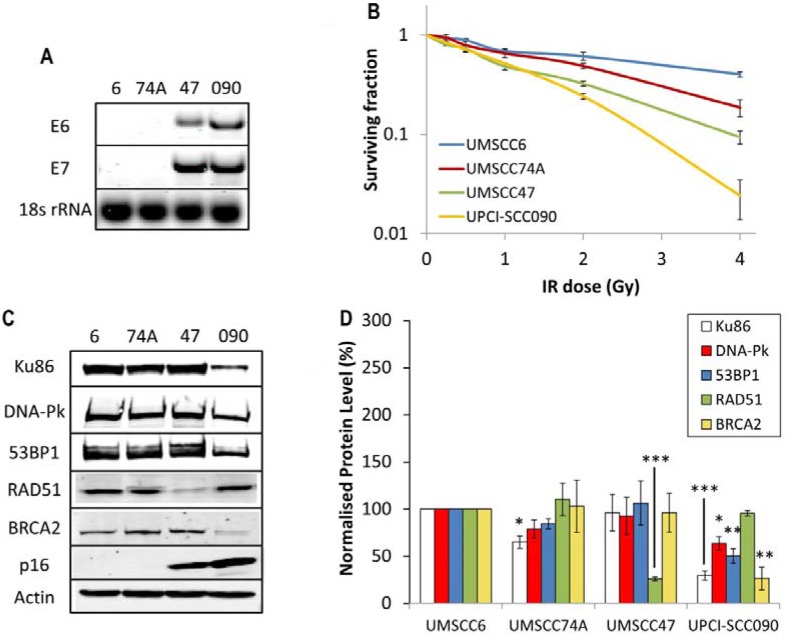
Figure 1. Analysis of radiosensitivity of HPV-negative and HPV-positive OPSCC cells and correlation with DSB repair protein levels.
(A) RT-PCR of cDNA prepared from OPSCC cells confirming HPV status through expression of E6 and E7 oncogenes, in comparison to 18s rRNA as a control, as analysed by agarose gel electrophoresis. (B) Clonogenic survival of OPSCC cells was analysed following treatment with increasing doses of x-ray irradiation (0–4 Gy). Shown is the surviving fraction with standard errors from at least three independent experiments. A comparison of the surviving fraction at 2 Gy (SF2) by one-way ANOVA reveals p < 0.01 (UMSCC6 vs UMSCC47), p < 0.005 (UMSCC6 vs UPCI-SCC090), p < 0.02 (UMSCC74A vs UMSCC47) and p < 0.002 (UMSCC74A vs UPCI-SCC090). (C) Whole cell extracts from OPSCC cells were prepared and analysed by 10 % or 6 % SDS-PAGE and immunoblotting with the indicated antibodies. (D) Levels of DSB repair proteins relative to actin were quantified from at least three independent experiments. Shown is the mean protein level relative to actin with standard errors from at least three independent experiments, normalised to those calculated in the HPV-negative UMSCC6 cell extracts which was set to 100 %. *p < 0.05, **p < 0.02, ***p < 0.005 as analysed by a one sample t-test of normalised protein levels in the respective cell extracts relative to the UMSCC6 extracts.

