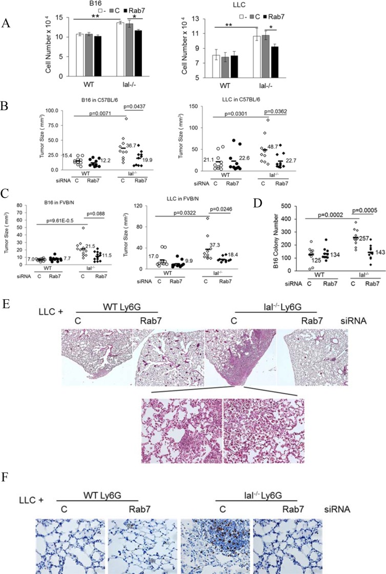
Figure 7. Rab7 GTPase controls tumor stimulation of lal−/− MDSCs.
(A) Ly6G+ cells from the wild type or lal−/− bone marrow were transfected with control or Rab7 GTPase siRNA for 24 h, and 2 × 10⁵ transfected Ly6G+ cells were co-cultured with B16 melanoma cells (2 × 10⁴) or LLC cells (2 × 10⁴) in vitro. Cells were counted 48 h later. Data were expressed as mean ± SD, n = 5. *P < 0.05, **P < 0.001; -, no transfection; C, transfected with control siRNA; Rab7, transfected with Rab7 siRNA. (B) Ly6G+ cells from the syngeneic C57BL/6 wild type or lal−/− bone marrow were transfected with control or Rab7 GTPase siRNA for 24 h. Transfected Ly6G+ cells were mixed with B16 melanoma cells (2 × 10⁵) or LLC cells (5 × 10⁵) (3:1), followed by subcutaneously flank injection into C57BL/6 wild type recipient mice. Tumor sizes were measured at post injection day 10. The average tumor size of each group and p values are marked with n = 10; (C) Ly6G+ cells from the allogeneic FVB/N wild type or lal−/− bone marrow were transfected with control or Rab7 GTPase siRNA for 24 h. Transfected Ly6G+ cells were mixed with B16 melanoma cells (2 × 10⁵) or LLC cells (5 × 10⁵) (3:1), followed by subcutaneously flank injection into the FVB/N wild type recipient mice. Tumor sizes were measured at post injection day 10. The average tumor size of each group and p values are marked with n = 12; (D) Ly6G+ cells from C57BL/6 wild type or lal−/− bone marrow were transfected with control or Rab7 GTPase siRNA for 24 h. Transfected Ly6G+ cells (2.5 × 10⁶) were mixed with B16 melanoma cells (5 × 10⁵) (5:1), followed by intravenously injection into C57BL/6 wild type recipient mice through tail vein. B16 melanoma colony numbers were counted at 2 weeks post injection. The average colony number of each group and p values are marked with n = 10; (E) Ly6G+ cells from C57BL/6 wild type or lal−/− bone marrow were transfected with control or Rab7 GTPase siRNA for 24 h. Transfected Ly6G+ cells (2.5 × 10⁶) were mixed with LLC cells (5 × 10⁵) (5:1), and intravenously injected into C57BL/6 wild type recipient mice through tail vein. Lung histology of LLC invasion was examined at 2 months post injection. The representative H & E sections were shown, n = 10 mice; (F) Ki67 staining of E. For A-F: -, no transfection; C, transfected with control siRNA; Rab7, transfected with Rab7 siRNA.

