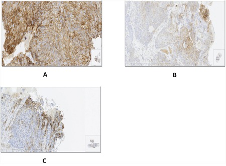
Figure 3. Different patterns of PD-L1 expression in ESCC specimens.

A. Diffuse PD-L1 expression in the presence of TILs; B. Regional expression of PD-L1 colocalized with TILs; C. PD-L1 expression at the invasive front.

A. Diffuse PD-L1 expression in the presence of TILs; B. Regional expression of PD-L1 colocalized with TILs; C. PD-L1 expression at the invasive front.