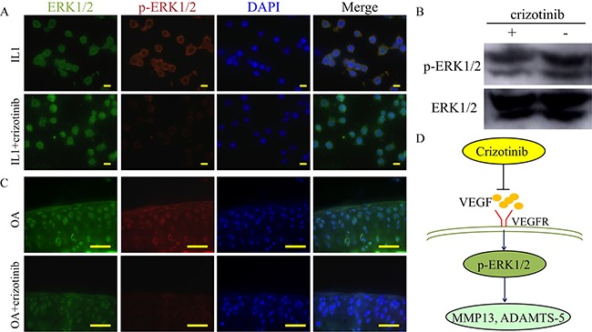
Figure 9. Mechanism of crizotinib in decreasing cartilage matrix degradation.

(A) Immunocytochemistry detection of ERK1/2 and p-ERK1/2 in mouse primary chondrocytes after stimulating with IL1β (10 ng/mL) with or without crizotinib (10 μM) for 1 h. Scale bars = 30 μm. (B) Western blot analysis of ERK1/2 signalling in mouse primary chondrocytes after IL1β treatment (cropped blots are displayed). (C) Immunocytochemistry was performed for ERK1/2 and p-ERK1/2 in OA mouse cartilage treated with or without crizotinib for 8 weeks. Scale bars = 100 μm. (D) A proposed model for the role of crizotinib in osteoarthritis treatment.
