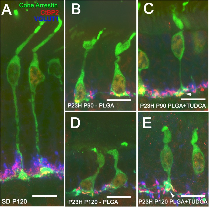
Fig 5. Photoreceptor morphology in vehicle- and TUDCA-PLGA-MSs-treated eyes.
Triple immunolabeling for cone arrestin (in green), VGLUT1 (in blue) and CtBP2 (in red) of retinal vertical sections from a P120 normal rat (Sprague Dawley, SD) (A) and P23H rats at P90 (B, C) and P120 (D, E), treated with unloaded PLGA MSs (B, D) or TUDCA-loaded PLGA MSs (C, E). Note that the typical cone pedicles (in green) containing synaptic vesicles (in blue) surrounding synaptic ribbons (in red) were less deteriorated in TUDCA-PLGA-MSs-treated P23H rats than in vehicle-treated animals. Scale bar, 10 μm.

