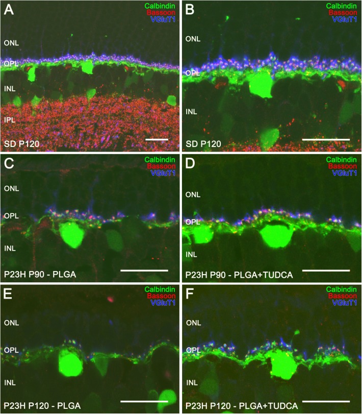
Fig 7. Horizontal cells and their synaptic contacts in vehicle- and TUDCA-PLGA-MSs-treated eyes.
Retinal vertical sections from a P120 normal rat (Sprague Dawley, SD) (A, B) and P23H rats at P90 (C, D) and P120 (E, F), treated with unloaded PLGA MSs (C, E) or TUDCA-loaded PLGA MSs (D, F). Horizontal cells were stained for calbindin (in green), synaptic ribbons were labeled using anti-Bassoon antibodies (in red) and synaptic vesicles in the photoreceptor were stained using antibodies against VGLUT1 (in blue). Note that pairings between photoreceptor axon terminals and horizontal cell dendrites were more numerous in TUDCA-PLGA-MSs-treated retinas, as compared to those observed in vehicle-treated retinas. ONL: outer nuclear layer, INL: inner nuclear layer, OPL: outer plexiform layer. Scale bar, 20 μm.

