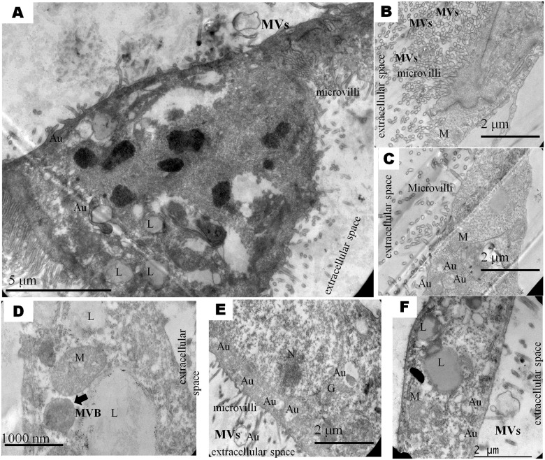
Fig 2. Representative microphotograph showing ultrastructure of bovine IVF and PA derived blastocysts.
Nucleus (N; in E), mitochondria (M; in C, D, F), lipids (L; in A, D, F), autophagosome (Au; in A, C, E, F), multivesicular body MVB (arrow in D) and microvilli (in A, B, C, D, E) shown superficially of the cell.

