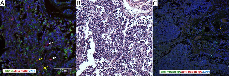
Fig 9. Immunohistochemical localization of ZIKV in fetal [and maternal] tissues.
(A) Immunofluorescent staining for ZIKV NS2B (red) and macrophage marker CD163 (green) in fetal axillary lymph node with a high vRNA burden. The white scale bar = 100 μm. (B) H&E stained near section of the tissue presented in 9A. (C) Nonspecific immunostaining with control isotypes for ZIKV NS2B and CD163.

