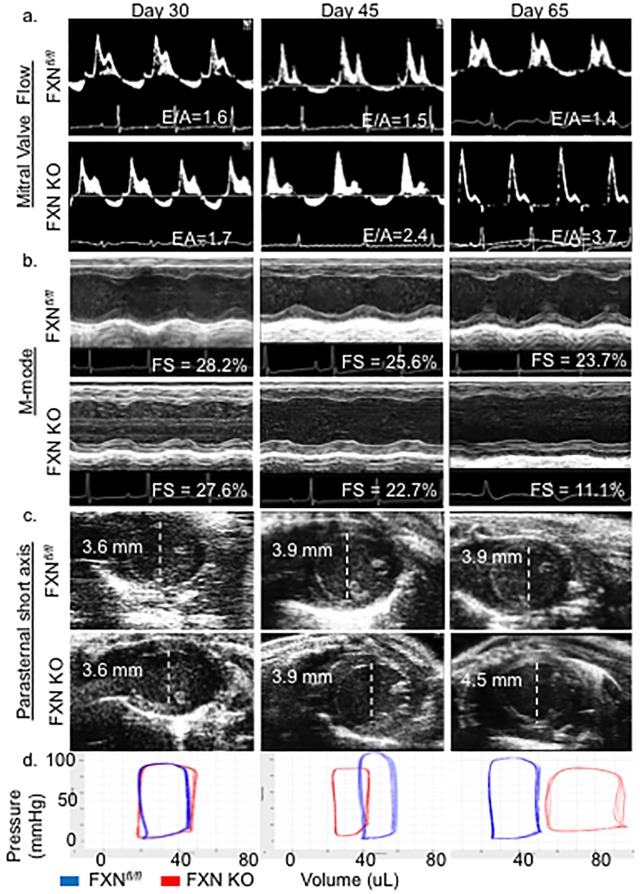
Fig 1. FXN KO mice exhibit diastolic dysfunction followed by dilated cardiomyopathy and heart failure.
(a) Representative mitral valve Doppler flow patterns (ratio of the early (E) to late (A) ventricular filling velocity, E/A) demonstrate restrictive cardiomyopathy in FXN KO at days of age 45 and 65. (b) Echocardiographic parasternal short axis M-mode images demonstrate progressive impairment in left ventricular wall movement with decreased fractional shortening (FS, %) in FXN KO. (c) Parasternal short axial images illustrate the transition to dilated cardiomyopathy with increased left ventricular internal diameter in diastole (LVIDd) in FXN KO at day 65. (d) FXN KO pressure volume loops represent increased end-diastolic volume (EDV) at day 65 compared to controls (p<0.001) with notable rightward shift of pressure-volume curves. Values indicated in (a), (b) and (c) are averages per group.

