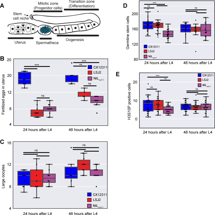
Fig 2. Analysis of components of egg-laying.
A. Schematic of the C. elegans gonad. Germline Stem Cells (not shown due to their large number) self-renew in the mitotic zone. As they migrate away from the stem cell niche, they undergo meiosis and differentiate into mature oocytes. Ovulation forces the primary oocyte into the spermatheca, which stores previously produced self-sperm, where it is fertilized and develops an eggshell. Fertilized eggs develop in the uterus until they are laid through the vulva. Only one of two gonads is shown. B. Number of fertilized eggs in the uterus as determined by DIC microscopy. C. Number of large oocytes as determined by DAPI staining and fluorescent microscopy. D. Number of germline progenitor cells. E. Number of cells undergoing mitosis in the mitotic zone, as determined by immunofluorescence to a post-translational modification (H3S10P) in Histone 3 correlated with chromatin condensation in mitosis.

