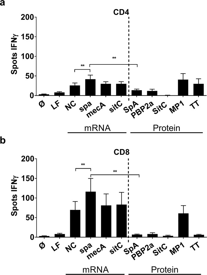
Fig 1. Comparison of IFNγ production by T cells stimulated with mRNA- or protein-derived staphylococcal antigens.
(a) CD4+ T cells and (b) CD8+ T cells in co-culture with MoDC were stimulated with either mRNA-encoded antigens or the corresponding protein antigens, e.g. spa / SpA, mecA / PBP2a and sitC / SitC. Lipofectamine (LF) alone, non-coding mRNA (NC) and a peptide pool from matrix protein 1 (MP1) of H1N1 Influenza virus and Tetanus toxoid (TT) served as controls. The number of IFNγ ELISpot spots after overnight culture is shown as mean values ± SEM of n = 8 donors. p**<0.01, p*< 0.05 (Wilcoxon matched-pairs signed rank test). Experiments were done in duplicates.

