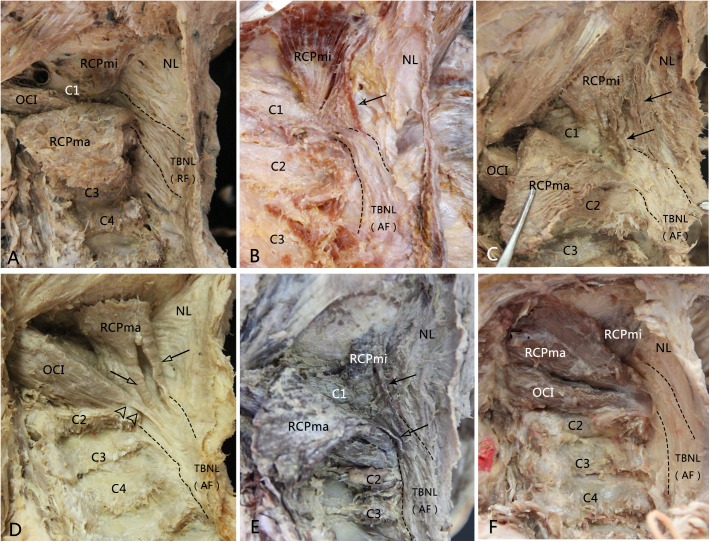
Fig 1. The second terminations of the suboccipital muscles (RCPmi, RCPma and OCI) and the types of the TBNL in the dissected specimens.
A, The TBNL within the NL was formed by radiate fibers. Opposite to the spinal processes of vertebrae C2 and C3, the radiate fibers originated from the posterior border of the upper part of NL, ran anterosuperiorly and straightly entered into the posterior atlanto-axial interspace. No second termination was found from the RCPmi, RCPma and OCI. B, The TBNL within the NL was formed by arcuate fibers. The arcuate fibers arose from the lower part of the posterior border of NL below the level of the spinal process of C3, ran anterosuperiorly, crossed over the spinous process of axis to continue into the posterior atlanto-axial interspace. A muscle bundle of the RCPmi separated and terminated at the level of atlanto-axial interspace. C, The RCPmi emitted multi-bundles of muscular fibers and attached to the arcuate TBNL at the level of the posterior arch of atlas and the axis. D, The TBNL was arcuate and the second terminations originated from the RCPma and the OCI. The OCI emitted a tendinous bundle which crossed behind the spinous process of the axis and continued with the arcuate TBNL. Two muscular bundles of the RCPma terminated at the TBNL at the level of the posterioratlanto-axial interspace. E, The TBNL was arcuate and the second terminations of the RCPmi and the RCPma were existed simultaneously. F, The arcuate TBNL was accompanied without a second termination of the suboccipital muscles. RF: radiate fibers; AF: arcuate fibers; Arrow: second termination of the RCPmi; Hollow arrow: second termination of the RCPma; Double hollow triangles: second termination of the OCI.

