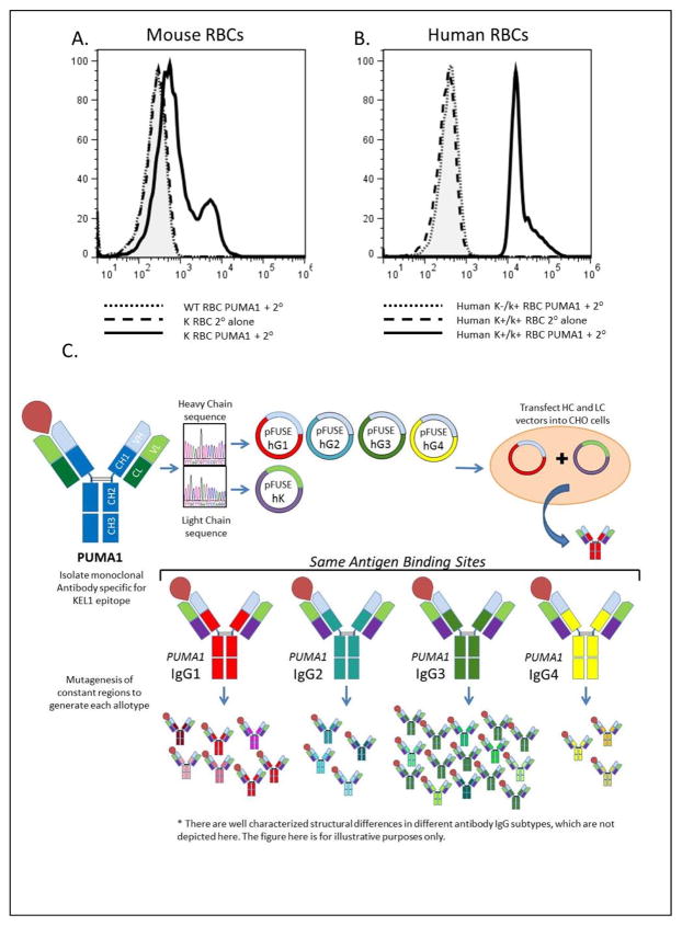
Figure 1.
Specificity of PUMA1 for the K antigen and strategy to generate PUMA1 variants. (A) Mouse RBCs were stained with PUMA1 antibody followed by secondary antibody (wild-type RBCs dotted line, K transgenic RBCs solid line). K transgenic RBCs were also stained with secondary antibody alone (dashed line). (B) Human RBCs with a K+k+ phenotype were stained with PUMA 1 followed by secondary antibody (solid line) or with secondary antibody alone (dashed line). RBCs with a K-k+ phenotype were stained with PUMA1 followed by secondary antibody (dotted line). (C) Diagram showing the general cloning and expression strategy.

