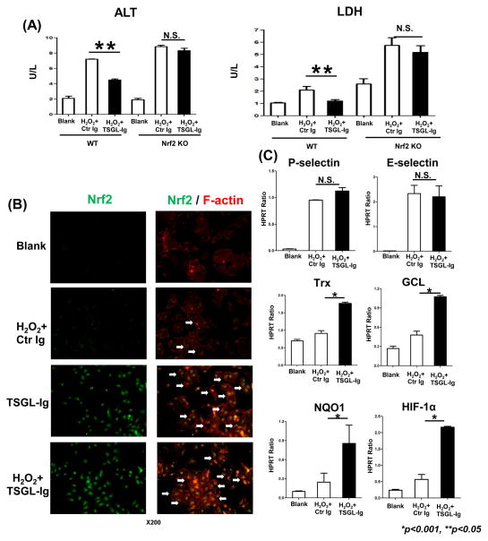
Figure 6.
The cytoprotective effects of TSGL-Ig upon LECs in vitro. Primary mouse liver endothelial cells from WT or Nrf2 KO donors were subjected to H2O2-stress with TSGL-Ig or control Ig supplement (20ug/ml at -1h) in 6h cultures. (A) ALT and LDH release; (B) Immunostaining of Nrf2 in LECs. White arrows indicate translocation of Nrf2 from cytoplasm to nucleus (Representative of n=3; magnification x200); (C) Quantitative RT-PCR-assisted detection of P-/E-selectin, Trx, GCL, NQO1, and HIF-1α. Data normalized to HPRT gene expression. Means ± SD are shown (**p<0.05, *p<0.001; n=4–6/group).

