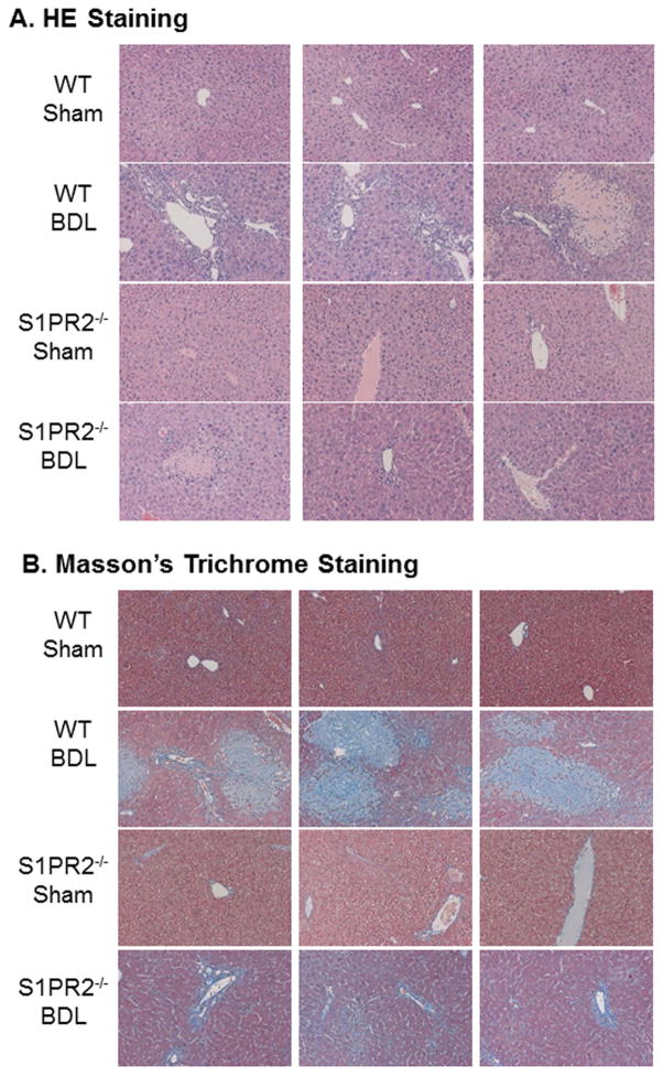
Figure 5. The role of S1PR2 in BDL-induced cholestatic liver injury.
Wild type and S1PR2−/− mice were subjected to 2-week BDL or sham operation as described in “Methods”. The liver sections were stained with H&E or Masson’s Trichrome. The images were taken with an Olympus microscope equipped with an image recorder using a 200 × lens. Representative images are shown. (A). H&E staining; (B) Masson’s Trichrome staining.

