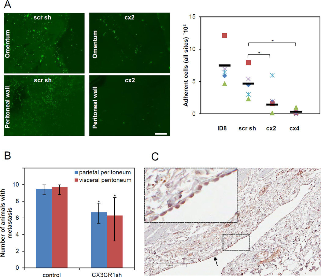
FIGURE 2. CX3CR1 supports peritoneal adhesion and tumor formation in a syngeneic model of ovarian carcinoma.
(A) Short-term in vivo adhesion assay demonstrates that CX3CR1 is important for peritoneal adhesion. Immunofluorescence images (green fluorescence) demonstrate cells adherent to omentum and peritoneal wall, as indicated, 4 hours following i.p. injection of the GFP-labeled control (scr sh group) and experimental (cx2 clone) ID8 cells into abdomens of C57BL/6 (5/group). Adherent cells were visualized under the fluorescence microscope in tissues excised from sacrificed control (injected with ID8 and scr sh) and experimental (injected with cx2 and cx4 clones) animals, enumerated using Zeiss AxioVert software, and plotted. Red squares – mouse #1, purple checks – mouse #2, blue flakes – mouse #3, green triangles – mouse #4, blue diamonds – mouse #5. Bars show an average number of adherent cells in each group. The differences between groups (cx2 vs scr sh and cx4 vs scr sh) were statistically analysed using both Mann-Whitney U test and Students’ t-test; * p<0.05. Differences between ID8 and scr sh groups were not statistically significant using both Mann-Whitney U test and Students’ t-test. All groups were statistically analysed using one-way ANOVA; p=0.0026. (B) Downregulation of CX3CR1 leads to significant reduction of tumor formation. The number of animals bearing metastasis on the organs covered by the parietal peritoneum and the visceral peritoneum in both control (ID8 and scr sh combined to a total of 20 animals; designated “control”) and experimental (clones cx2, cx3, and cx6 combined to a total of 30 animals; designated “CX3CR1sh”) groups was calculated and plotted. Animals were considered positive for metastasis at each site if lesions visible with the naked eye were present upon dissection when animals reached humane endpoints. The data are shown as average ± standard deviation and were statistically analyzed using Students’ t-test.; * p<0.05. (C) Normal human mesothelium is strongly CX3CL1-positive. Expression of CX3CL1 in human mesothelium was determined using immunohistochemistry and anti-CX3CL1-specific antibodies. A typical image of the CX3CL1 immunostaining in normal mesothelium from intestine/colon of a 55 year old woman is shown (core B1, Supplementary Table 1). Black arrow points to the mesothelial monolayer. Brown, CX3CL1; blue, hematoxylin. Bar, 200 micron. Insert is a 5-fold magnification of the area outlined with dashed lines.

