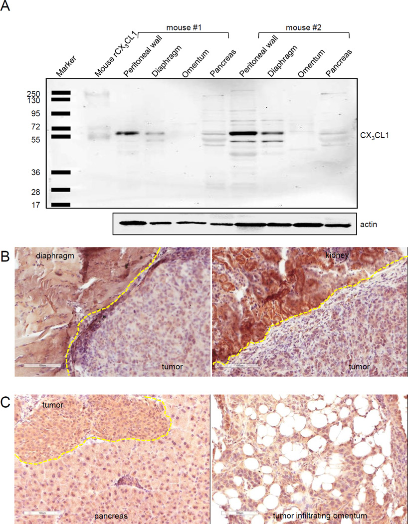
FIGURE 6. Role of tumoral and stromal CX3CL1 in CX3CR1-dependent metastatic dissemination.
(A) Expression of CX3CL1 in specimens of tumor-naive mouse omentum, pancreas, peritoneal wall, and diaphragm. Expression of CX3CL1 in total cell lysates (10 µg protein/lane) of peritoneal wall, diaphragm, omentum, and pancreas obtained from tumor-naive C57BL/6 mice (n=2) was detected using Western blot. Left lane shows positions of the molecular weight marker proteins. Mouse recombinant CX3CL1 (10 ng) served as a positive control. Actin served as a loading control. (B,C) Tumoral and stromal CX3CL1 expression. CX3CL1 in tumor and invaded stroma, as specified for each host site, was visualized with immunostaining in organ sites dissemination to which correlated with CX3CR1 expression in tumor cells (diaphragm, kidney, (B)) and where it did not depend on CX3CR1 expression in the tumor cells (pancreas, omentum, (C)). Images were generated using Aperio ScanScope; bar, 100 micron. Dashed yellow lines separate tumors from stroma. Brown, CX3CL1; blue, hematoxylin.

