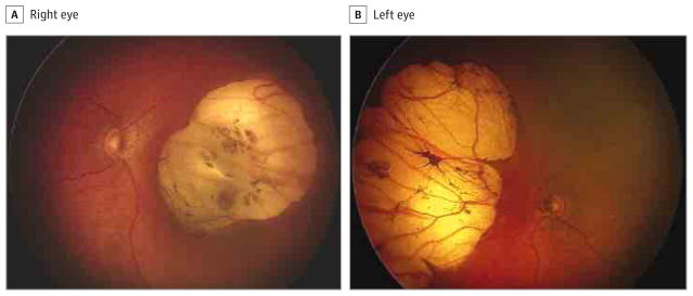
Figure 5. Fundus Photographs of a 20-Day-Old Infant.
The right eye has optic disc hypoplasia, peripapillary nasal atrophy, and an excavated nasal round lesion with a hyperpigmented halo, with a colobomatous-like aspect (A), and the left eye has optic disc hypoplasia, peripapillary nasal atrophy, and a retinal nasal lesion with a similar pattern (B).

