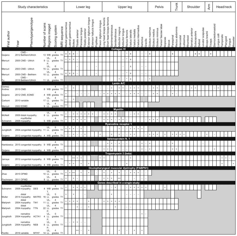
Table 6.
Patterns of muscle involvement and sparing in MRI studies describing congenital myopathy, congenital muscular dystrophy (CMD), Emery–Dreifuss muscular dystrophy (EDMD), distal myopathy, and oculopharyngeal muscular dystrophy (OPMD)
Shaded boxes represent muscles that were not scanned or analyzed. A * denotes fat replacement in the center of the muscle. A ± denotes fat replacement at the muscle periphery
T trunk, P pelvis, UL upper leg, LL lower leg, WB whole-body

