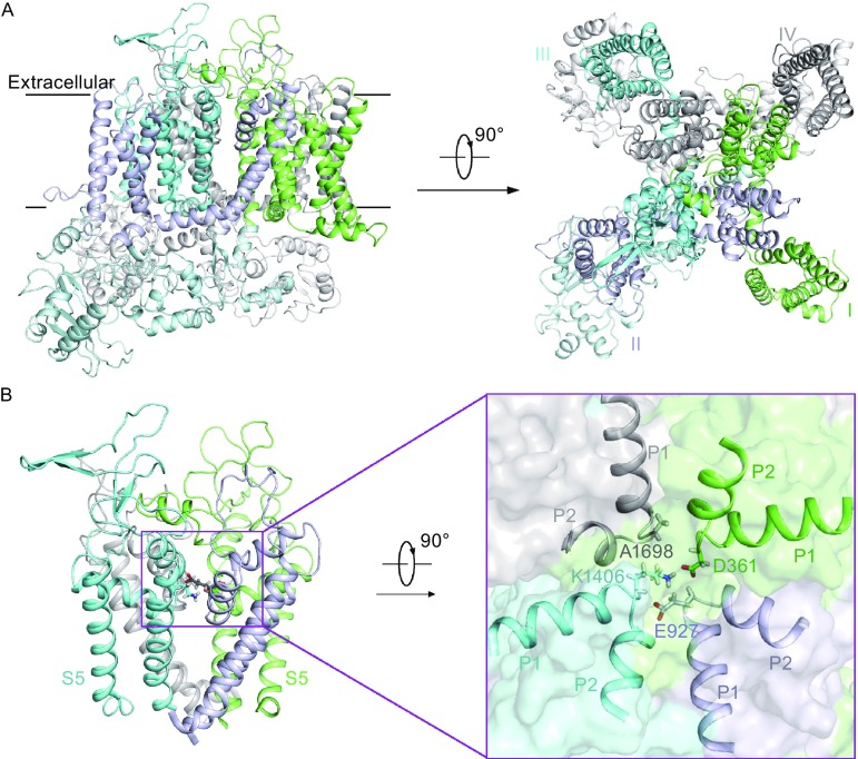
Figure 1.
Homology model structure of human Nav1.7 sodium channel. (A) Intra-membrane view and extracellular view of the structure model of Nav1.7. The four domains are colored green, light blue, cyan, and gray for domain I, II, III, and IV, respectively. (B) The pore domain of Nav1.7 structure model. The S5–S6 segments of Nav1.7 are shown and the four selectivity filter amino acids are shown as sticks (left). A close-up view of the four SF residues, D361 in domain I, E927 in domain II, K1406 in domain III, and A1698 in domain IV (right)

