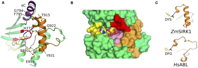
FIGURE 3.
ZmSIRK1 activation segment is highly structured. (A) Cartoon representation of ZmSIRK1 around the activation segment. Residues taking part in the stabilization of this segment are shown in stick. Possible hydrogen bonds are represented as dashed lines. (B) Surface representation of ZmSIRK1 nucleotide-binding site showing that the protein activation segment (orange) occludes the region normally available to interact with the target peptide. AMP-PNP is shown as spheres. (C) The 310 helix immediately C-terminal of ZmSIRK DYS motif (top) is also seen in inactive state mammalian kinase domains (bottom – human ABL; PDB ID 2G1T). The Cαs from putative phosphorylation sites in ZmSIRK1 and from known phosphorylation sites in HsABL are shown as spheres.

