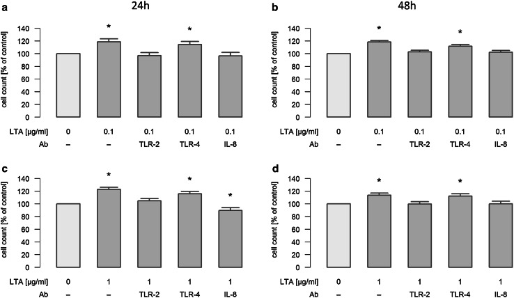
Fig. 4.
Mechanisms of LTA-induced proliferation of A549 cells. A549 cells were either sham-incubated (control) or exposed to 0.1 (a/b) or 1 µg/ml (c/d) of LTA for 24 or 48 h in the absence or presence of neutralizing antibodies targeting TLR-2, TLR-4 or IL-8. A549 proliferation was assessed by automatic cell counting. All data are expressed as percentage of baseline proliferation of sham-incubated cells, which was set to 100%. Means ± SEM of at least four independent experiments are given. Values marked with an asterisk differ significantly from controls (p < 0.05)

