Fig.1.

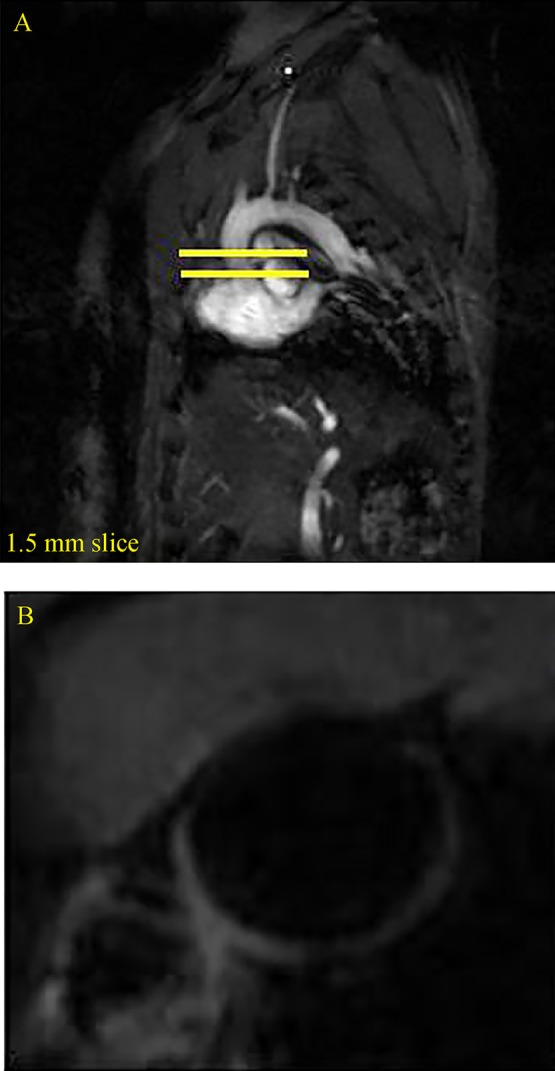
MRI images in sagittal view of the vasculature that were used to identify the anatomy and define the imaging places (A) and MRI images of aortic arch after treatment with LDE-paclitaxel (B).

MRI images in sagittal view of the vasculature that were used to identify the anatomy and define the imaging places (A) and MRI images of aortic arch after treatment with LDE-paclitaxel (B).