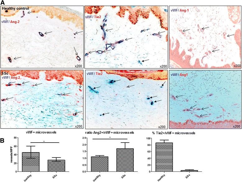
Fig. 2.

Expression of Ang/Tie2 in dermal microvessels of SSc patients and controls. a As assessed by double (+/+) staining of skin biopsies, Ang-2 (red staining) was more abundantly expressed in dermal microvessels (vWF+; blue staining) of SSc patients compared with healthy controls, whereas Ang-1 (red staining) did not differ. The expression of the membrane-bound Tie2 receptor (red staining) was remarkably reduced in dermal microvessels (vWF+; blue staining) of SSc patients compared with healthy controls. Vessels are indicated by arrows. b shows the respective semi-quantitative analyses of dermal Ang-2 and mbTie2 expression as well as the reduced microvascular density of SSc patients compared with healthy controls. Pictures are representative examples of 24 SSc patients and 19 healthy controls. Ang Angiopoietin, SSc systemic sclerosis, vWF von Willebrand factor
