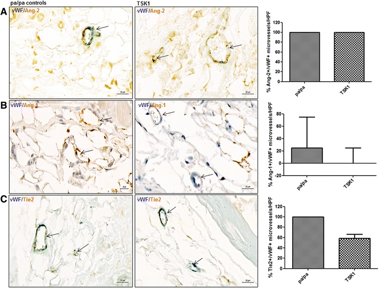
Fig. 6.

Expression of Ang/Tie2 in dermal microvessels of TSK1 mice. a, b, c In the TSK 1 model, no significant changes could be observed with respect to the expression of Ang-2, Ang-2, Tie2 (brown staining) in microvessels (vWF positive; blue staining) in the hypodermis. Vessels are indicated by arrows. Pictures are representative examples of four TSK mice and four pa/pa controls. Ang Angiopoietin, HPF high power field, TSK1 tight skin 1, vWF von Willebrand factor
