
Fig. 1.
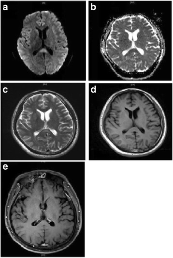
Initial cranial MRI of the patient. The lesion in the midline of SCC was hyperintensity on DWI (a) and T2WI (c), decreased ADC value (b), isointense signals on T1WI (d), and no contrast enhancement (e)
Official websites use .gov
A
.gov website belongs to an official
government organization in the United States.
Secure .gov websites use HTTPS
A lock (
) or https:// means you've safely
connected to the .gov website. Share sensitive
information only on official, secure websites.
Initial cranial MRI of the patient. The lesion in the midline of SCC was hyperintensity on DWI (a) and T2WI (c), decreased ADC value (b), isointense signals on T1WI (d), and no contrast enhancement (e)