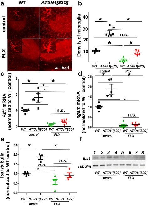
Fig. 1.

PLX treatment depletes microglia in the cerebella of ATXN1[82Q] mice. ATXN1[82Q] and wild-type (WT) mice were fed PLX3397(PLX) or regular (control) chow from 3 weeks of age. At 3 months of age, the cerebella was dissected and used for immunofluorescence (IFC) or RNA analysis. a Representative confocal images of 3-month-old mice stained with a microglia specific Iba-1-antibody. Insets in each image demonstrate hypermorphy of PLX-treated surviving microglia. Scale bar, 100 μm. b Density of Iba1 positive microglia in the cerebellar cortex (N = 5 per each genotype). c, d Cerebellar RNA was used for quantitative RT-PCR with primers for microglia specific genes Aif1(c, N ≥ 4) and Itgam (d, N ≥ 3). e, f Western blotting for Iba-1 demonstrates decrease in PLX-treated mice. (f, N ≥ 3). Each dot represents one mouse, and values indicate mean ± SEM. Asterisk indicates p < 0.05 by one-way ANOVA followed by Bonferroni post hoc test, and n.s. indicates that results are not statistically different (p > 0.05)
