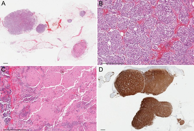
Figure 5.
Histopathologic images. (A) Low-power view showing multiple tumor deposits in adipose tissue. Hematoxylin & eosin, 2×. (B) Each tumor deposit is composed of nests of cells ranging from oval to spindle, without overt cytologic atypia or significant mitotic activity. Hematoxylin & eosin, 10×. (C) There is focal necrosis in a few tumor deposits. Hematoxylin & eosin, 10×. (D) The tumor cells are strongly and diffusely positive for synaptophysin. Immunoperoxidase, 2×. Bar, 1 mm.

 This work is licensed under a