Fig. 3.

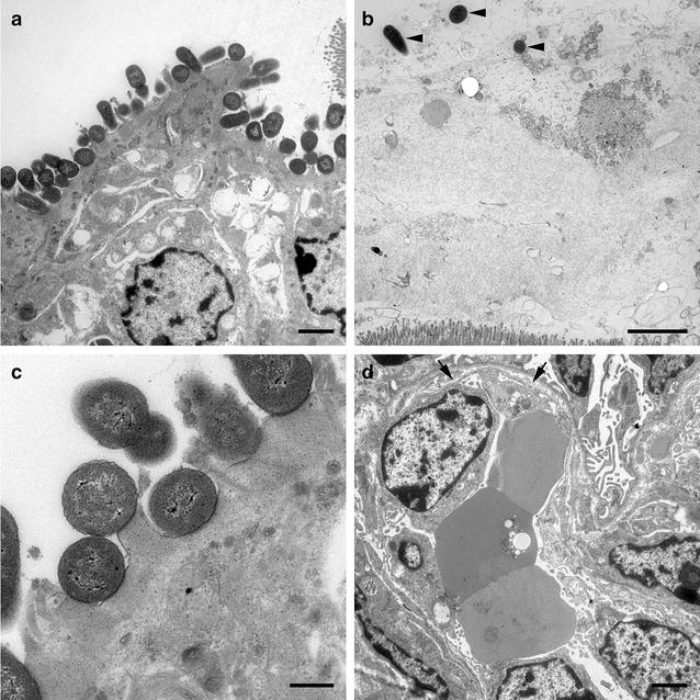
Electron microscopic images from E. coli O157:H7 and E. coli O104:H4 infected animals, respectively. a Colon ascendens of piglet infected with E. coli O157:H7. Bacteria are in intimate contact to epithelial cells, causing A/E lesions. Bar = 2500 nm. b Colon ascendens of piglet infected with E. coli O104:H4. Scattered bacteria in mucus above epithelial layer can be seen (arrowheads). No direct contact to epithelial cells is determinable. Bar = 2500 nm. c Colon ascendens of piglet infected with E. coli O157:H7. Bacteria are in intimate contact to epithelial cells, causing A/E lesions. Bar = 500 nm. d Glomerulum of the kidney of an E. coli O157:H7 infected piglet. Dilated subendothelial space caused by detachment of endothelial cells from basement membrane (arrow). Bar = 2500 nm
