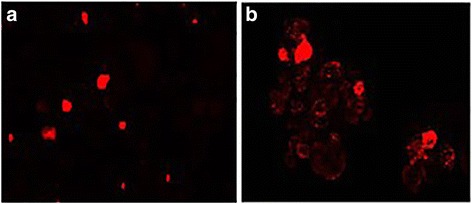
Fig. 2.

Images of PKH-26 staining during spheroid formation (magnification 100X). Single cells labeled with PKH-26, derived from mechanic dissociation of OVA-BS4 spheroids; day 0 (a). After 4 days, most of the cells underwent PKH-26 dilution in the newly formed spheroids while few retained the red label (b)
