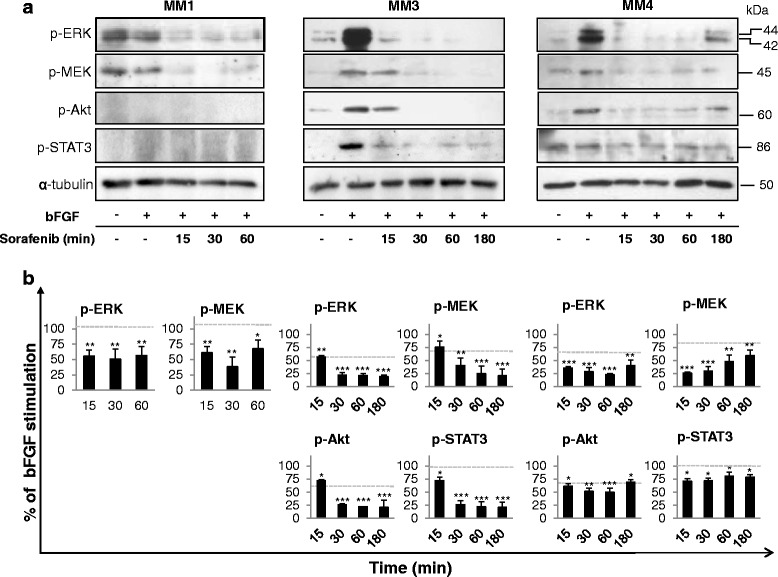
Fig. 6.

Sorafenib suppresses intracellular signaling in bFGF-stimulated MPM TICs. a Western blot analysis using antibodies specific to the phosphorylated proteins in MM1, MM3, and MM4 cells treated with IC50 sorafenib and stimulated with 20 ng/ml bFGF for the indicated time points (15–180 min). Representative immunoblots are reported. b Densitometric scanning of the immunoreactive bands followed by correction for α-tubulin loading control, from independent experiments. Values expressed as a percentage of the value obtained for the bFGF-stimulated cells in the same blot. Results are mean ± SEM of three experiments. *p <0.05, **p <0.01, ***p <0.001. Dotted lines, basal (untreated) levels. bFGF basic fibroblast growth factor
