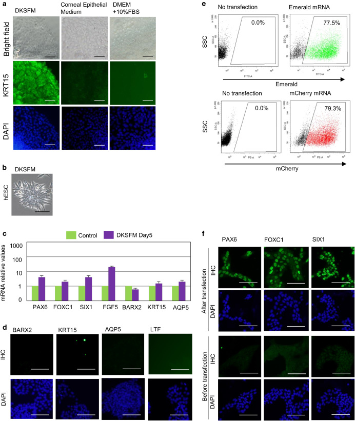
Fig. 2.
Establishment of a culture condition for the overexpression of transcription factors. a Immunohistological analysis of mouse E16.5 LGE in DKSFM (left), corneal epithelial medium (center), and DMEM with serum (right). Scale bar 100 µm. b Phase-contrast images of hESCs in DKSFM at day 5. Scale bar 100 µm. c Relative mRNA expression profiles in hESCs after culture in DKSFM and the control cells that also grown for the same length of time (5 days) in basal media as the experimental cells. Error bars represent mean ± standard deviation (SD) of three samples. d Immunohistochemical analysis of the cultured hESCs in DKSFM with antibodies against BARX2, KRT15, AQP5, LTF at Day 5. Scale bar 100 µm. e The analysis of expression rate of GFP and mCherry proteins by modified mRNA transfection into hESCs using FACS analysis. f PAX6, FOXC1, and SIX1 expression in cells 8 h after transfection with the synthetic modified mRNAs

