
Fig. (1).
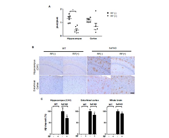
Chronic RF-EMF exposure reduces Aβ deposition in Tg-5xFAD mice. A. Aβ in Tg-5xFAD and WT mice at the age of 9.5 months following RF-EMF exposure for 8 months. The Aβ42 and Aβ40 in the brain lysate of Tg-5xFAD mice were measured by color metric ELISA assay. The graph shows a ratio of Aβ42 peptide and Aβ40 in lysates of the hippocampus and entorhinal cortex. B. Representative images of Aβ deposition in the CA1 region of the hippocampus and entorhinal cortex of Tg-5xFAD and WT mice. Brown spots indicate Aβ plaques. Scale bar: 100μm. C. Quantification of Aβ deposition in hippocampus, entorhinal cortex and whole brain area. Data indicate arbitrary value compared to control Tg-5xFAD mice. Values are presented as the mean ± SEM (n=6). Statistical differences were established by two-tailed t-test analysis for comparison between 5xFAD-RF(-) and Tg-5xFAD-RF (+) for p-value less than 0.05.