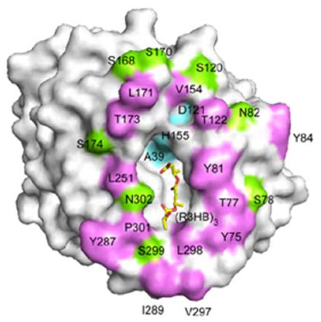
Figure 15.
A molecular surface representation of the PHA depolymerase from P. funiculosum centering on the mouth of the crevice. Positions of solvent-exposed hydrophobic residues (purple), as well as polar (green) and catalytic triad (cyan) residues, are indicated. A model of the 3HB trimer bound in the crevice is shown as a yellow stick model [86]. Reproduced with permission from Hisano et al., J. Mol. Biol.; published by Elsevier, 2006.

