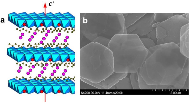
Figure 1.
(a) Schematic illustration of the layered double hydroxide (LDH) structure showing the metal hydroxide octahedra stacked along the crystallographic c axis (indicated as a red arrow). Water (grey) and anions (pink) are present in the interlayer region. The green parts correspond to MII cations and the red dots to MIII cations. (b) Scanning electron microscope (SEM) image of typical LDH crystals.

