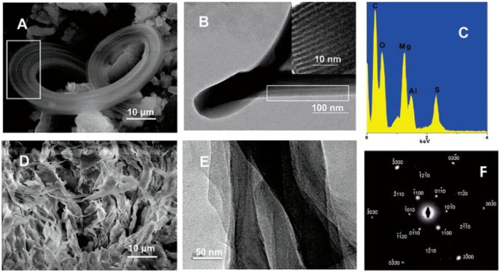
Figure 4.
(A) SEM image of a belt-like LDH structure with the parallel linear pattern highlighted in the white rectangle. (B) TEM image of one belt reveals the lamellar structure; a HRTEM image of this structure is shown in the inset, and (C) spectrum of the chemical compositions analyzed by energy dispersive X-ray (EDX). The belts were easily exfoliated to give structures shown by the SEM image in (D) and TEM image in (E). (F) The corresponding selective area electron diffraction pattern. Reprinted with permission from [19], © American Chemical Society, 2005.

