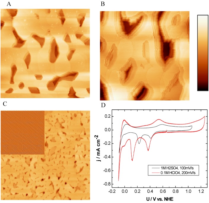
Figure 2.
EC-STM, SECPM, AFM and CV of Ru(0001): (A) EC-STM (500 nm × 500 nm, hmax = 12.17 nm), US = 500mV vs. NHE, (B) SECPM (500 nm × 500 nm, hmax = 17.22 nm) image of Ru(0001) in 0.1 M HClO4 at US = 500 mV vs. NHE, (C) Contact mode AFM in air (5 µm × 5 µm, hmax = 40 nm, Inset: atomic resolution, 12 nm × 12 nm) and (D) CVs obtained in 1 M H2SO4 (black curve) and 0.1 M HClO4 (red curve) with a scan rate of 100 mV∙s-1 and 200 mVs-1, respectively. Imaging conditions: STM: IT = 1 nA, Ubias = 100 mV, SECPM: ∆U = 5 mV.

