Figure 10.
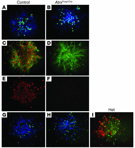
Increased apoptosis but normal proliferation in ATRX-deficient primary cortical cultures. (A–I) Primary cultures of cortical progenitor cells were established from E12.5 telencephalon of AtrxFoxg1Cre mice (B, D, F, and H), control Cre– littermates (A, C, E, G), or heterozygote (Het) Cre+ littermates (I), and were grown for 6 days in culture. (A and B) BrdU staining for 16 hours of control (A) and knockout (B) colonies demonstrates that the proliferative capacity of the cultured cells is not diminished in the absence of ATRX protein expression. (C and D) Control progenitors show a high level of ATRX protein (red) in differentiating MAP2-positive cells (green). AtrxFoxg1Cre progenitors that are ATRX deficient (lack of red staining in D) still express high levels of MAP2. (E and F) Corresponding ATRX staining of clones in G and H demonstrates the absence of ATRX protein in AtrxFoxg1Cre progenitors. (G and H) Merged images of TUNEL-positive cells (green) and DAPI staining (blue). Note the increased level of TUNEL staining in AtrxFoxg1Cre cell colonies after 6 days in culture. (I) Heterozygous Cre+ progenitor cell clone stained for ATRX protein (red) and TUNEL (green), clearly showing that ATRX-deficient areas of the clone display higher levels of apoptosis. Magnification, ×20 (A–I).

