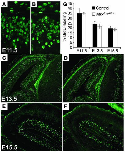
Figure 7.
Normal proliferation in AtrxFoxg1Cre cortex during embryogenesis. Timed matings were set up between homozygous Atrx-floxed female mice and Foxg1Cre-heterozygous males. Pregnant female mice were subjected to a 1-hour BrdU pulse prior to sacrifice. Embryos were fixed and frozen, and sections were processed for BrdU immunostaining at E11.5 (A and B), E13.5 (C and D), E15.5 (E and F). Normal controls (A, C, and E) are compared with AtrxFoxg1Cre mice (B, D, and F). (G) BrdU-positive cells were counted in an identically sized area and data are expressed as a percentage of the total number of cells (DAPI-positive) at E11.5, E13.5, and E15.5. No statistically significant differences in proliferation were observed at any of the time points examined (E11.5, n = 4, P = 1.000; E13.5, n = 6, P = 0.181; E15.5, n = 5, P = 0.482). Magnification, ×40 (A and B), ×20 (E and F), and ×10 (C and D).

