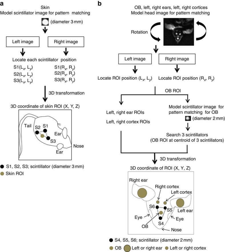
Figure 2. Determination of ROI positions in six areas of freely moving mice.
First, the frames with OB, skin, ear, cortex and three scintillators on each image are selected by ‘Mouse Tracker'. (a) For skin, three scintillator ROIs are identified using a model scintillator image. The skin ROI was assumed to be at the centre of the baseline of the triangle formed by the three scintillators. (b) For OB, ear and cortex ROIs, a model head image is used as a search model for pattern matching in all frames of the video with any angle of rotated versions of the model. Ear and cortex ROIs are defined relative to the top-left corner of the model head image. The OB ROI was assumed to be at the centroid of the triangle formed by the three scintillators. Next, 3D coordinates of ROI in each area are computed by transformation matrix (see the ‘Methods' section for details). 3D coordinates of ROIs are illustrated in the skin and head of the mouse.

