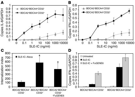
Figure 6.
CD32+ PDCs internalize and respond to SLE-ICs. (A and B) PDCs from normal donors were isolated on a MoFlo cell sorter using fluorescently labeled antibodies against BDCA2, BDCA4, and CD32. The CD32– and CD32+ PDC subsets were stimulated with the indicated concentrations of SLE-ICs. Cells were harvested at 3 and 8 hours, and expression of IL-8 (A) and IFN-α (B) was determined by QPCR. (C) Alexa Fluor 633–conjugated SLE-ICs in the presence or absence of 10 μl FuGENE6 were incubated with CD32+ and CD32– PDCs at a ratio of 10:1 for 15 minutes at 37–C. Internalization was measured by fluorescent microscopy. Data are presented as the number of internalized complexes per 100 cells × 100. *P < 0.01 versus control, Student’s t test. Error bars indicate standard deviation of triplicate samples. (D) Total RNA was isolated from PDCs stimulated with 100 ng/ml SLE-ICs in the presence or absence of 10 μl of FuGENE6. Expression of IFN-α was determined by QPCR and is depicted as the number of copies of mRNA per copies of the control mRNA GAPDH.

