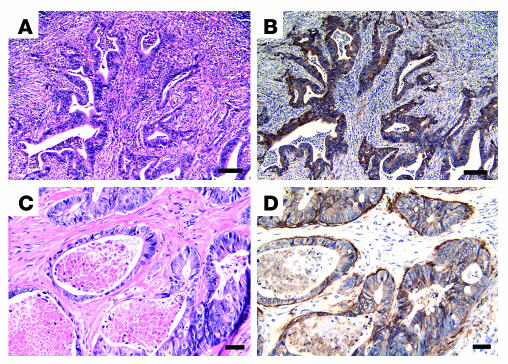
Figure 8.
In vivo αvβ6 protein expression in human colorectal metastases. Representative H&E (A and C) or β6 immunohistochemistry (B and D) in lymph node (A and B) or liver tissue (C and D) containing human colorectal metastases. β6 Immunoreactivity in both samples is restricted to the metastasized tumor cells. Scale bars: 100 μm (A and B) and 50 μm (C and D).

