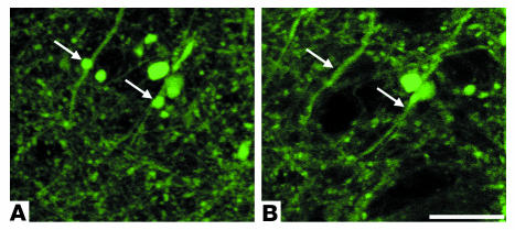
Figure 4.
Morphological recovery of amyloid-associated neuritic dystrophy in a PDAPP;Thy1:YFP double-transgenic mouse after anti-Aβ antibody (10D5) treatment. The 2 panels show the same population of YFP-labeled neurites, which are associated with a neuritic plaque imaged on the initial day of surgery (A) and 72 hours later (B). The antibody was administered directly to the surface of the brain during the cranial-window surgical procedure on day 0. The arrows in the day 0 image (A) point to 2 enlarged dystrophic neurites, which are absent in the day 3 image (B). The associated amyloid is not shown. Scale bar: 20 μm.

