
Extended Data Figure 6. Characterization of chromothripsis and polyploidy in metastases.
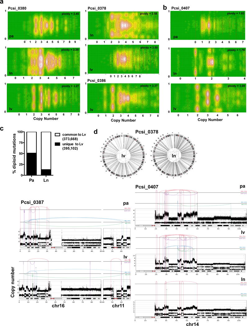
(a) CELLULOID plots illustrating polyploidy in metastases. In Pcsi_0380, the primary tumour was directly available for analysis. Similarly to Pcsi_0378, multiple metastases were polyploid suggesting primary tumour was also polyploid. The primary tumour was unavailable for sequencing in this case. (b) A case (Pcsi_0407) with discordant ploidy amongst different metastases. (c) Percent of diploid mutations from liver metastases that are shared (white) or unique (black) in comparison to the primary tumour or the lymph node metastasis. (d) Plots of chromothripsis events in metastases. pa – primary tumour; lv – liver; ln – lymph node.