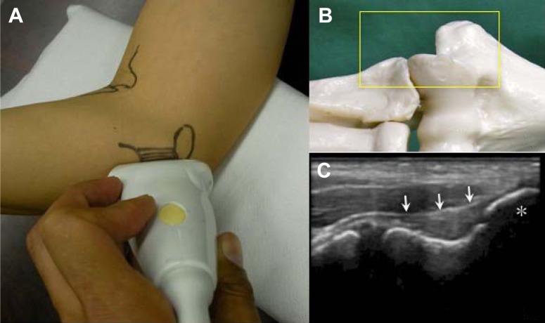
Figure 1.
Ultrasonographic examination of the medial elbow. The elbow was flexed 90° and the forearm was placed in the supinated position. (A) A linear transducer was placed on the medial aspect of the elbow to obtain an image that included (B) the top of the medial epicondyle (MEC) (*), the anterior bundle of the medial ulnar collateral ligament (MUCL), and the sublime tubercle. (C) The MUCL (arrow) was identified as a band-like structure that attached to the MEC and sublime tubercle.

