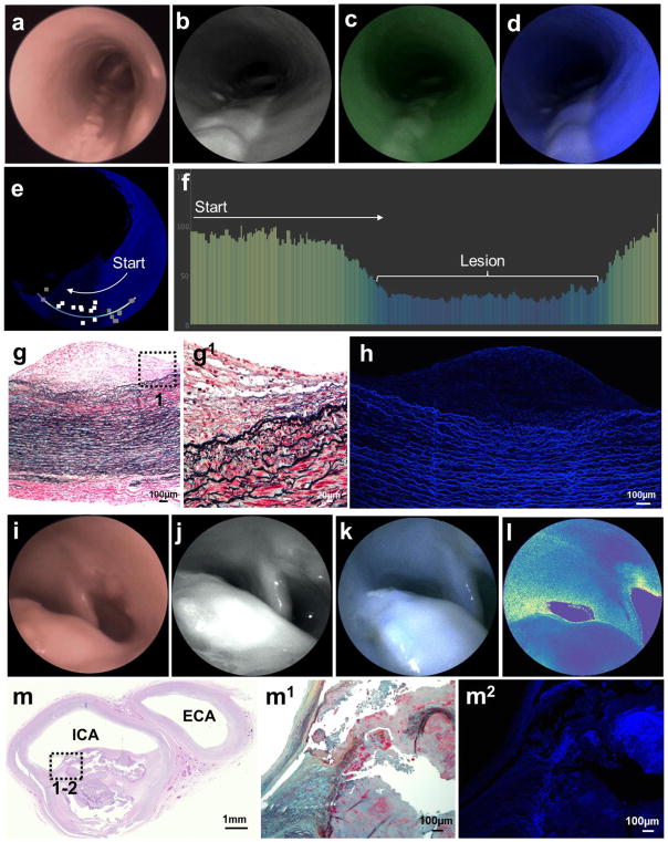
Figure 3. Multimodal endovascular scanning fiber angiography of (a–h) intermediate and (i–m) advanced lesions.
a&i, White-light endoscopic system images. b&j, Scanning fiber endoscopy images with red reflectance. c, Scanning fiber endoscopy images with red reflectance and laser-induced green fluorescence. d&k, Scanning fiber endoscopy images with red reflectance and laser-induced blue fluorescence. e, Distance-compensated fluorescence image. a–e, Intermediate Lesion: Note the large longitudinal elevated lesions with loss of blue and green autofluorescence, which is better appreciated in the distance-compensated fluorescence image. Note the 10 random square areas in lesion and 10 in background used for target-to-background ratio. f, Color-coded bar chart of pixel values of a line traced in (e) reveals >50 points drop of autofluorescence intensity registered from the lesion in comparison to surrounding vascular background, on average 2.5 times stronger. g, Histological analysis of arterial cross-section reveals multiple layers of fat-laden macrophages at the intima with sparse free-lipid deposits in deep portion of the lesion (Movat’s stain). This is a Grade III intermediate lesion. g1, Higher magnification of the plaque shoulder [dotted box in (g)] shows no distinct necrotic core. h, Confocal microscopy revealed very low autofluorescence derived from the intermediate atheroma, compared to the intense autofluorescence originating from elastin in the tunica media. i–l, Advanced Lesion: Protruding lesion with smooth homogeneous surface in carotid bifurcation causing significant luminal stenosis. l, This lesion is associated with low autofluorescence, which is better appreciated in distance-compensated pseudocolor map. m, Histological analysis of arterial cross-section reveals a fibroatheroma (Grade IV) extending from carotid bifurcation into proximal internal carotid artery [(m) hematoxylin and eosin stain, with dotted insert represented in (m1), Movat’s pentachrome stain]. m2, Confocal microscopy shows a poorly fluorescent collagenous fibrous cap overlying a necrotic core with heterogeneous fluorescence.

