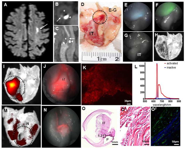
Figure 5. Multimodal endovascular scanning fiber angioscopy of proteolytic activity in endarterectomy specimen.
An elderly man suffered a left hemispheric ischemic stroke [(a) brain MRI; arrow points to ischemic region] secondary to ipsilateral complicated carotid plaque [(b) computed tomographic angiography of axial plane, and (c) sagittal reconstruction with arrows pointing to the remaining vascular lumen, arrowheads pointing to calcification, and asterisks indicating fibroatheroma]. d, At surgery, the fibroatheroma was removed and found to have a ruptured cap associated with a large intraluminal thrombus. e–f, Scanning fiber endoscopy imaging of the region highlighted in (d) revealed delicate blue and green autofluorescence originating from the fibrous cap, with preserved reflectance and absent autofluorescence from the intraluminal thrombus. g, No fluorescent signal was identified in the reflectance or red channel, which was then confirmed by (h) scanning tissue with IVIS. i, After incubation with MMPSense, strong signal is observed from the cap surrounding the intraluminal thrombus using IVIS, which is clearly detected by (j) scanning fiber angioscopy with high spatial resolution. Note the geographic and intensity differences of specific red signals obtained from the (i–j) endoluminal surface in comparison with (m–n) the abluminal surface of the plaque. k, Red signal detected with scanning fiber angioscopy from activation of the molecular probe was confirmed with upright confocal microscopy in fresh full-mount specimen. l, Point-spectrometer revealed a maximum fluorescence emission of 666nm for activated MMPSense 645, and inactive MMPSense 645 was optically silent. o, Histological analysis of the specimen revealed a large intraluminal thrombus with an underlying complicated fibroatheroma. p, In situ zymography of the fibrous cap underlying the thrombus with (q) DQ-gelatin-confirmed high gelatinase activity from matrix metalloproteinases.

