. 2017 May 8;6:e24494. doi: 10.7554/eLife.24494

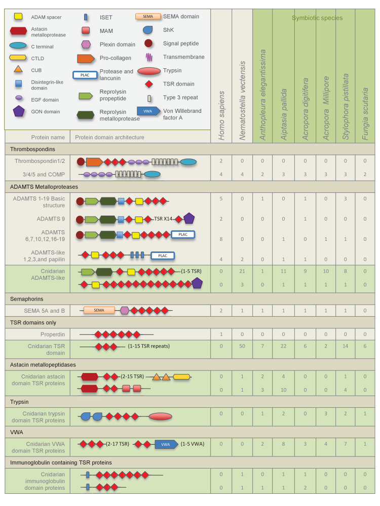
Figure 2. Domain architecture of cnidarian TSR super-family proteins compared to known vertebrate TSR-domain-containing proteins.

DOI: http://dx.doi.org/10.7554/eLife.24494.004

Figure 2.

Figure 2—figure supplement 1. The TSR domain is very well conserved from cnidarians up to humans, with binding motifs for glycosaminoglycans (GAGs) and the type B scavenger receptors, CD36/SRB1.

Figure 2—figure supplement 1.

All three-dimensional folding sites are present as described by Tan et al. (2002) for the crystal structure of human TSP1 TSR2. Six conserved cysteine residues are highlighted in yellow and form three disulfide bridges (C1–C5, C2–C6 and C3–C4). Three conserved tryptophan residues are shown in blue boxes and mark the ‘WXXW’ protein-binding motif. Amino acids that form the R layers are marked with purple boxes, and pairings forming 3 R layers are as follows: R3-R4, R2-R5 and R1-R6. The Β strands are annotated at the bottom in blue strands A, B and C. Please refer to Tan et al. (2002) for a more detailed explanation of the three-dimensional folding.