
Figure 2. MSCs inhibit differentiation of myeloid progenitor cells in vitro.
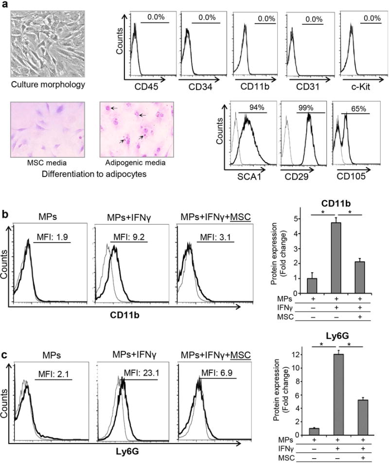
A. Left Panel: Expansion and characterization of MSCs. Microscopic images of MSCs cultured in MSC or adipogenic media (×40 magnification). Oil-Red-O staining after 2 weeks showed red colored fat vacuoles (black arrows) in the cytoplasm of MSCs cultured in adipogenic media, confirming their differentiation into adipocytes. Right Panel: Representative flow cytometry plots demonstrating the phenotype of bone marrow derived MSCs as CD45−CD34−CD11b−c-Kit−CD31−Sca-1+CD29+CD105+ cells. B. Representative flow cytometric histograms and bar chart demonstrating CD11b expression by myeloid progenitors (MPs) cultured with or without MSCs with IFNγ stimulation for 72 hours. C. Representative histograms of flow cytometric data and bar chart showing Ly6G expression by myeloid progenitors (MPs) cultured with or without MSCs with IFNγ stimulation for 72 hours. Results are representative of 3 independent experiments. Myeloid progenitors were isolated from a pool of 5–6 animals in each experiment. P values are calculated using student’s t-test and data is represented as mean ± SEM. *p< 0.0001.